🧠 Imagine your brain surgeon has never practiced on a tumor like yours before. In neurosurgery, a surgeon's only chance to experience a specific complex tumor has always been during the actual operation — a single, unrepeatable moment. Now, a Japanese university-industry collaboration is changing that forever with a 3D model that lets surgeons practice on your exact tumor — again and again.

The Surgeon's Dilemma: No Practice Allowed

Surgeons, like athletes, need practice. But in neurosurgery, there has been a fundamental problem that has persisted for decades: you simply cannot rehearse on the real thing.

Every brain tumor is unique. Its shape, size, location, and relationship with surrounding nerves and blood vessels differ from patient to patient. Yet traditionally, a neurosurgeon's only opportunity to navigate a specific tumor's complexity was during the actual surgery itself. For the patient on the operating table, it was always the surgeon's first and last attempt at that particular case.

Surgeons can study CT and MRI scans beforehand, of course. But the information gleaned from a flat screen is fundamentally different from the three-dimensional tactile reality of tissue, bone, and surgical instruments. Textbooks and monitors simply cannot convey the spatial relationships between a tumor and the delicate structures surrounding it.

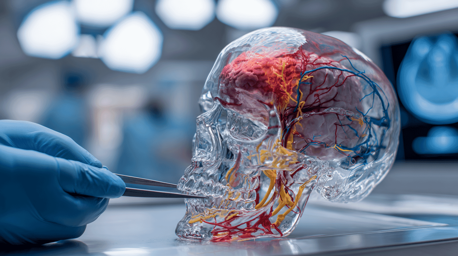
Enter the "Hakata Model"

In February 2025, Kyushu University's Graduate School of Medical Sciences and Japan Medical Company announced the launch of a joint research project to tackle this problem head-on. Their creation is called the "Hakata Model" — a next-generation medical training platform named after the historic district of Fukuoka, the city where Kyushu University is located.

The Hakata Model's core innovation lies in its ability to reproduce a specific patient's intracranial anatomy, including their unique tumor, with extremely high precision based on CT/MRI data. This is not a generic skull model. It faithfully recreates the tumor's shape, extent, and its spatial relationship with surrounding nerves and blood vessels for each individual case — a truly "patient-specific" training tool.

The joint research involves Professor Koji Yoshimoto and Lecturer Taisuke Kuga from the Department of Neurosurgery at Kyushu University's Graduate School of Medical Sciences. The project builds on the KEZLEX precision medical model technology that Japan Medical Company has refined over approximately 25 years.

KEZLEX: 25 Years of Building Models You Can Actually Drill

The foundation of the Hakata Model is KEZLEX, a precision medical replica model that reproduces the internal and external structure of human bones from 3D CT/MRI data. Its defining feature is that it can actually be drilled — it replicates the tactile sensation of cutting into real bone, and even the feel of applying surgical clips.

Japan Medical Company's predecessor, Ohno Kogyo, was founded in 1897 as a steel trading company. In 1999, the company began researching 3D printing technology for medical applications and succeeded in reproducing the stapes — the smallest bone in the human body. This technology has been patented internationally and is now used in over 50 countries, including the United States, Europe, China, and Southeast Asia.

Until now, KEZLEX has primarily been used to reproduce anatomical structures. The Hakata Model adds a crucial new element: the individual tumor itself. Integrating anatomical models with tumor models has the potential to fundamentally transform the concept of surgical training.

From "Once Only" to "As Many Times as Needed"

The essential transformation the Hakata Model brings is converting a "one-time surgical experience" into "shareable, repeatable training." Several applications are envisioned.

First, repetitive skill practice. Surgeons can grasp the three-dimensional spatial relationship between the tumor and surrounding nerves and blood vessels before surgery, then practice the required techniques as many times as needed. The specific hand movements that previously existed only in a veteran surgeon's muscle memory can now be experienced by junior doctors.

Second, location-independent training. Because the model is physical and portable, training is not limited to the operating room. The same model can be shipped to regional hospitals or overseas institutions, potentially narrowing the educational gap between urban medical centers and rural facilities.

Third, enhanced team-based medicine. Specialists in microscopic surgery, endoscopic surgery, and exoscopic surgery can gather around the same model to discuss and compare different approaches. This enables teams to collaboratively develop optimal surgical strategies that minimize complication risks.

Professor Yoshimoto noted that the model allows surgeons to visualize tumor shapes and nerve-vessel relationships unique to individual cases, enabling risk prediction. This underscores the Hakata Model's role not just as a training tool but as a surgical planning aid.

3D-Printed Surgical Models: A Global Movement

The application of 3D printing to surgical training is accelerating worldwide. Since the first skull model was created from CT images in 1990, the 2010s saw costs drop to practical levels, with research advancing in spinal surgery, vascular surgery, and brain tumor surgery.

However, current 3D-printed models share a common limitation: haptic realism remains insufficient. While bone texture can be replicated, the elasticity of brain tissue and the softness of tumors are not yet fully reproducible. Typical 3D-printed surgical models cost between $100 and $1,000, though real costs including labor are substantially higher.

What sets the Hakata Model apart is the level of tactile fidelity achieved through 25-plus years of KEZLEX refinement. Rather than simply reproducing shapes, the focus on replicating the feel that a surgeon's hands experience reflects a distinctly Japanese monozukuri (craftsman-like manufacturing) philosophy. This meticulous approach to manufacturing could set a new benchmark in the field.

Patient Safety and the Future of Medicine

The Hakata Model's development ultimately serves patient safety. When surgeons can adequately practice before an operation, their ability to handle unexpected situations improves and complication risks decrease.

Furthermore, the ability to accumulate and share valuable surgical cases as "educational assets" will accelerate the transfer of expertise. Knowledge and experience that once lived exclusively in a veteran surgeon's hands can now be accessed by the next generation of doctors.

Kyushu University and Japan Medical Company have set their sights on establishing the Hakata Model as an internationally standardized education and evaluation platform originating from Japan. How the fusion of Japanese precision manufacturing and medical research will reshape neurosurgical education worldwide remains a story worth following.

How are surgeons trained in your country? What do you think about using 3D-printed models to practice brain surgery? We'd love to hear your perspective.

References

Reactions in Japan

The Hakata Model news is blowing up in our resident LINE group chat. Honestly, if our generation gets to use this, the number of skull base approach practice reps will be on a completely different level. Being able to re-experience our attending's 'that one case' is huge.

I agree 0
I disagree 0

My father had brain tumor surgery, so I'm truly grateful for this initiative. I always wanted to ask 'Doctor, how many times have you done this surgery?' but never could. Knowing surgeons can practice on a model first gives a completely different level of reassurance.

I agree 0
I disagree 0

KEZLEX's 'drillable model' is technically quite advanced. They replicate bone hardness through resin formulation, but integrating soft tissue tumors into that must make material selection extremely challenging. Looking forward to follow-up reports.

I agree 0
I disagree 0

The concept of 'making a one-time surgical experience repeatable' is intuitively compelling. But cost per model and delivery time will be key to widespread adoption. I'd also like to see a roadmap from research stage to clinical implementation.

I agree 0
I disagree 0

At a regional core hospital like ours, we only get a few complex cases per year. Not being able to build experience like urban university hospitals has always been a concern. If the Hakata Model lets us practice urban cases in rural settings, it could elevate regional healthcare.

I agree 0
I disagree 0

During my clinical clerkship in neurosurgery, the doctor explained the risk of damaging a certain nerve given the tumor's position, but looking at CT images alone didn't really click. Being able to see it on a physical model would probably change my level of understanding entirely.

I agree 0
I disagree 0

Great example of industry-academia collaboration between Kyushu Uni and a company. But university research often ends at the announcement stage, so I hope they follow through to commercialization. Japan is said to be strong in basic research but weak at what comes after.

I agree 0
I disagree 0

In preop conferences we discuss approach risks, but having a physical model might also help nurses better visualize intraoperative preparation. It would be amazing if the whole team, not just doctors, could use it for practice.

I agree 0
I disagree 0

Curious how this will coexist with VR simulation. Tactile feedback is still overwhelmingly better with physical models, but with haptic technology advancing, who knows what things look like in 5-10 years. I think using both together is the best approach.

I agree 0
I disagree 0

Wonder if this will be covered by insurance. If training costs get folded into surgery fees, medical costs rise. But if complications decrease and hospital stays shorten, the total could go down. I hope they pursue health economics evaluation too.

I agree 0
I disagree 0

Love the name 'Hakata Model' lol. Hakata ramen, Hakata Torimon sweets, and now Hakata Model. Another thing for Fukuoka to be proud of.

I agree 0
I disagree 0

I had my brain tumor surgery 5 years ago. Only afterward did I realize the immense pressure my surgeon was under. If technology like this reduces surgeons' psychological burden and improves patient safety, it deserves more support.

I agree 0
I disagree 0

Interesting initiative, but wouldn't shifting to robotic surgery like da Vinci be a more fundamental solution than increasing practice reps with 3D models? An approach that eliminates hand tremor entirely seems more impactful long-term.

I agree 0
I disagree 0

I've used KEZLEX at conference hands-on sessions before, and the drilling feel is indeed excellent. But whether they can reproduce tumor tactile sensation is unknown. The real technical challenge will be expressing infiltrative tumors with ambiguous borders with brain tissue.

I agree 0
I disagree 0

If it's based on KEZLEX which is already in 50 countries, the Hakata Model has a foundation for international expansion. But FDA clearance and regulatory approval in each country is a separate challenge. For educational use, device approval may not be needed, but it depends on distribution strategy.

I agree 0
I disagree 0

This model could be used for preoperative patient explanations too. Many patients don't grasp CT cross-sections. Showing them in 3D — 'Your tumor is here, and we'll approach from here' — would dramatically improve understanding and consent quality.

I agree 0
I disagree 0

Voices from Around the World

Dr. Marcus Chen

We use 3D-printed models at Stanford too, but I haven't seen an approach that integrates tumor and anatomy while replicating tactile feedback. The 25-year refinement behind KEZLEX is a serious advantage. Would love to try this in our lab.

Sarah Mitchell

Under the NHS training system, surgical training hours keep getting cut. Models like this could help increase the density of limited training time. My concern is the implementation cost in the UK though.

Prof. Hans-Werner Müller

At Charité Berlin, we're also developing brain tumor surgery simulators, but haptic realism is the biggest challenge. We've tried hybrid models using calf brain, but an industrial approach like KEZLEX is fascinating.

Rajesh Kapoor

In India, patient numbers vastly exceed available neurosurgeons. If training tools like this become affordably available, they could revolutionize education for our rapidly growing pool of young surgeons. It all depends on cost.

Dr. Isabelle Fontaine

In France, cadaver-based hands-on training is the norm, but there are ongoing ethical debates. 3D models avoid ethical issues entirely and can reproduce patient-specific cases. I'd attend a presentation on this at a Paris conference.

James O'Brien

In a vast country like Australia, it's tough for rural hospital surgeons to access the same training as urban counterparts. If you can just ship a model for training, it could bridge the education gap in remote medicine.

Kim Soo-jin

Samsung Medical Center in Korea is also advancing 3D-printed skull base model research. Japan-Korea collaboration would be interesting. Developing models optimized for Asian cranial morphology benefits both countries.

Carlos Mendoza

Surgical simulation equipment in Mexican public hospitals is extremely limited. If they developed an affordable version, it could hugely benefit medical education in developing countries. Maybe Japan's ODA programs could help introduce it.

Dr. Emily Zhao

China's 3D-printed surgical model startups are multiplying, but I have to admit Japan leads in haptic replication accuracy. However, cheaper Chinese models have the scalability advantage. Competition seems inevitable.

Michael Torres

Honestly, I think VR combined with AI has more future potential. Physical models cost time and money per unit, but VR can generate infinite cases from data alone. Just waiting for haptics technology to catch up.

Dr. Amara Okafor

Nigeria faces a severe shortage of neurosurgeons — fewer than 100 specialists for 200 million people. What matters is whether educational tools like this reach developing nations. I hope Japanese technology contributes to closing the global healthcare gap.

Anna Kowalski

I coordinate neurosurgery training at a Polish university hospital, and securing cadavers gets harder each year. Reusable high-precision models would help both ethically and logistically. Would love to know pricing.

David Park

Japan's monozukuri spirit is alive in the medical world again. Not just robots, but this kind of painstaking model technology that improves surgical safety — it's so Japanese. Quiet but innovative.

Dr. Fatima Al-Rashid

Saudi Arabia is rapidly modernizing healthcare infrastructure. Advanced educational tools like this align with Vision 2030's healthcare goals. We're interested in technology transfer from Japan.

Lucas Schmidt

We're validating 3D-printed neuroendoscopy simulators in Basel, with material costs around $67-$112. If the Hakata Model's tactile accuracy can be achieved at this price range, it's transformative. But 25 years of tech refinement likely means a premium price.

Patricia Souza

Under Brazil's SUS public health system, surgical wait lists are long, and it takes time for young surgeons to accumulate enough case experience. If simulation shortens the learning curve, it could indirectly reduce wait times too.