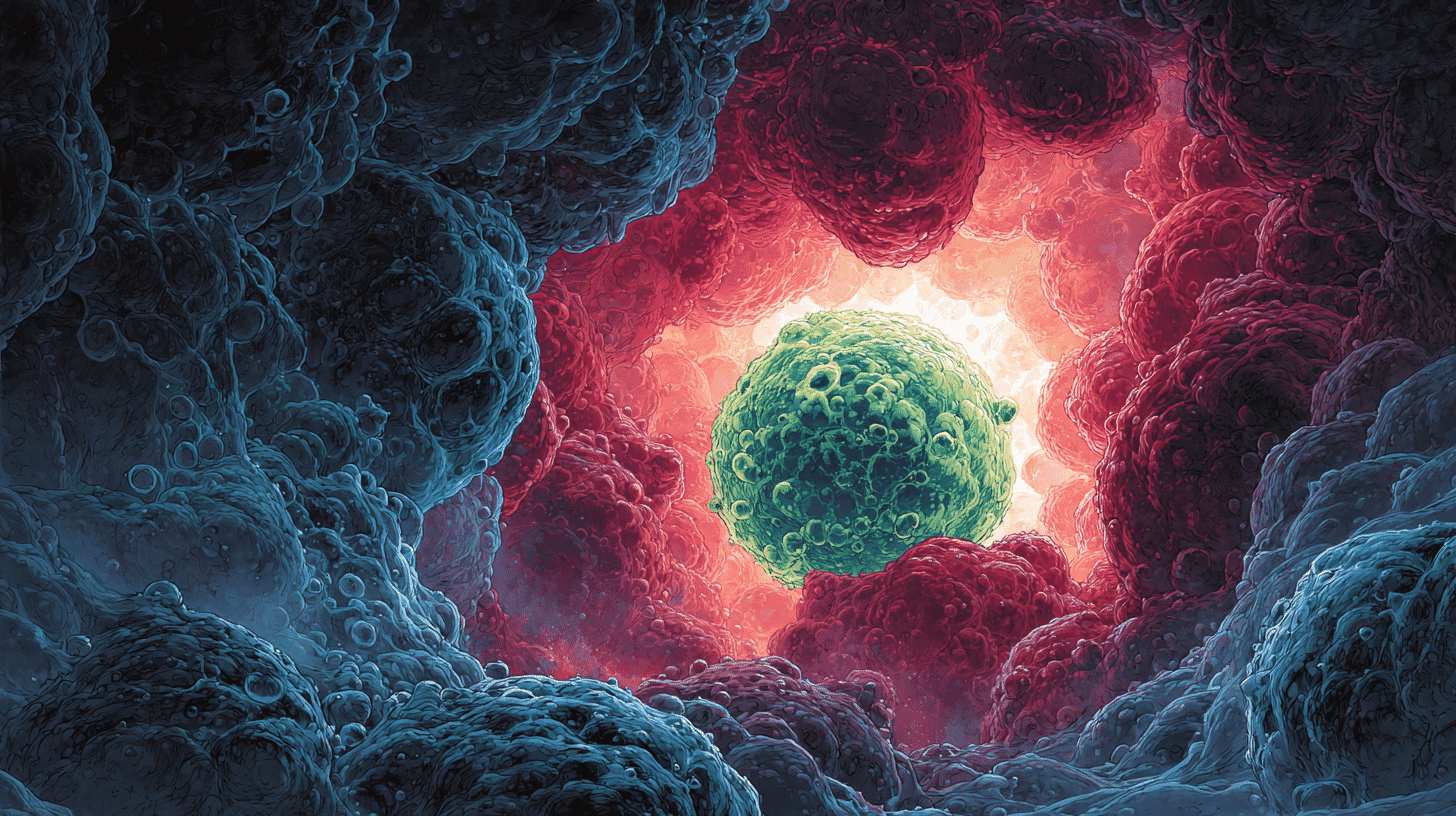
🧬 Imagine a pig that grows a kidney made from your own cells, ready to transplant, with little risk of rejection. For more than fifteen years, one stubborn mystery has blocked that idea: the instant scientists tried to grow one animal's cells inside another, the cells disappeared. A team in Tokyo and at Stanford has finally caught the culprit in the act, and it was hiding in plain sight. The host embryo's very first immune cells were eating the foreign cells alive.
The discovery, published online in the journal Cell on June 5, 2026, gives a name to a problem that has quietly limited regenerative medicine for over a decade: xenophagocytosis, roughly "eating cells from another species."
The organ you grow to order
Start with the shortage, because that is what makes any of this matter. In the United States alone, organdonor.gov counts more than 103,000 people on the transplant waiting list, with about 17 dying every day before an organ arrives. Someone new is added every eight minutes or so. Japan's situation is even tighter, with one of the lowest donor rates among wealthy nations.
For years, the lab of Hiromitsu Nakauchi, who now splits his work between the Institute of Science Tokyo (formed in 2024 by the merger of Tokyo Tech and Tokyo Medical and Dental University) and Stanford, has chased a radical answer. The idea is called blastocyst complementation, and it is simpler than it sounds. Take a host embryo that has been engineered so it cannot build a particular organ, say a pancreas. Inject donor stem cells. The donor cells rush to fill the empty space, the "organ niche," and build the missing organ almost entirely out of themselves.
It is not a hypothesis. In 2010, Nakauchi's team grew a rat pancreas inside a mouse, though it came out mouse-sized, too small to yield enough islets to treat anything. In 2017 they ran the experiment in reverse, injecting mouse stem cells into rat embryos to grow rat-sized mouse pancreases. Islets taken from those and transplanted into diabetic mice held blood sugar normal for more than 370 days, with immunosuppressing drugs given only for the first five days after surgery and none thereafter.
That last detail is the whole point. This is not the same as the pig-organ transplants that have made headlines recently, where a whole, gene-edited pig kidney is sewn into a human and the patient takes lifelong drugs to keep their immune system from destroying it. The dream here is different: an organ built from the patient's own cells, grown inside an animal that simply provides the construction site. Your cells, animal scaffolding, no rejection.
The wall between species
There was always a catch. Grow mouse cells in a mouse and the donor cells thrive. Grow rat cells in a mouse, crossing the species line, and they contribute far less. Try human cells, and the drop-off is steeper still. Scientists called it the xenogeneic barrier, and for fifteen years its cause stayed, in the researchers' own framing, elusive and multifactorial.
The leading suspects were developmental. Different species grow at different speeds; their cells carry slightly incompatible adhesion molecules; maybe the foreign cells simply couldn't keep up. Plausible, but never quite the full story. Something was deleting the donor cells, and no one had pinned down what.
A coincidence in the calendar
The break came from looking at the clock instead of the cells.
Nakauchi's team noticed that rat donor cells in a mouse embryo did not fade gradually. They crashed during a narrow window: embryonic days 9.5 to 11.5. When the researchers asked what else happens in that exact window, the answer was striking: it is precisely when the host's first immune cells, called primitive macrophages, appear. This is long before the adaptive immune system, the T cells and B cells we usually blame for rejection, even exists.
So they watched. Under the microscope, host macrophages were swallowing donor cells whole. And here is the unsettling part: most of those donor cells had not died first. They were not apoptotic debris being tidied away. They were alive, healthy, and being eaten anyway. The team named the phenomenon xenophagocytosis.
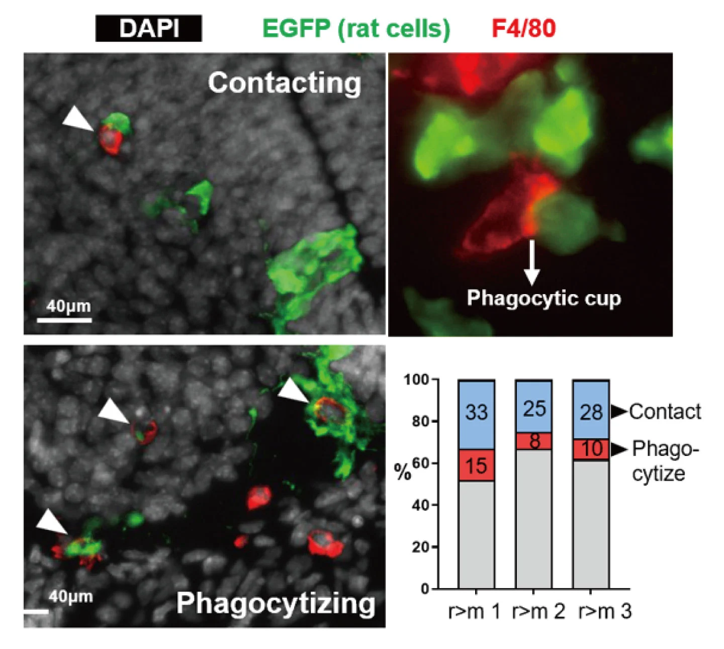
Source: Institute of Science Tokyo (Science Tokyo)
A name tag that reads "eat me"
Why would an immune cell devour a perfectly living one? The team traced the trigger to a kind of molecular name tag.
In the foreign environment, donor cells are under constant stress. That stress raises the calcium level inside them, which switches off an enzyme called a flippase (ATP11C) whose job is to keep the cell membrane organized. With the flippase down, a lipid called phosphatidylserine, normally tucked safely on the inner face of the membrane, flips to the outside surface. On a cell, an exposed patch of phosphatidylserine is a universal "eat me" signal. The macrophage reads it through a receptor called Axl, and begins to feed.
If that eat-me / don't-eat-me logic sounds familiar, it should. One of the study's co-authors, Stanford's Irving Weissman, helped pioneer it in cancer research, where blocking a tumor's "don't eat me" signal, CD47, unleashes the immune system to attack it. Here the team ran the same idea in reverse: instead of stripping a "don't eat me" tag away, they wanted to hand one to the donor cells.
Three ways past the guard
Once the mechanism was clear, the fixes followed almost logically. The researchers developed three approaches: remove the host's macrophages (or knock out their Axl receptor) so there is no one to do the eating; give donor cells the "don't eat me" tag CD47, borrowing straight from the cancer playbook; or boost the flippase so the "eat me" tag never surfaces in the first place.
Each one sharply raised donor-cell survival. In embryos where host macrophages were genetically ablated, rat contribution reached as high as 77 percent in the lung and 57 percent in the heart. Combined with macrophage removal, the success rate for growing a rat pancreas inside a mouse climbed. Most important for the long game, when the team removed host macrophages from mouse embryos carrying human cells, the human cells survived better too, strong evidence that the same guard stands watch across species, including ours.
How far this actually gets us
It is worth being honest about the distance still to travel. This work lives in mice and rats, plus human cells grown inside mouse embryos. Human cells still contribute only a tiny fraction of the embryo proper, about one cell in 2,500 at day 11.5, though in the yolk sac that figure reached as high as 20 percent. The pig, the host everyone actually has in mind for human organs, is the next test, not a finished one. No one is growing a human kidney in a barn yet.
What changed is the map. For years the species barrier was treated as a developmental puzzle; this study reframes a central piece of it as an immune one, with a specific mechanism and three concrete ways around it. The same insight may even feed back into the pig-organ transplants in the news, since macrophages turn out to be part of that fight as well.
And then there is the harder question, the one no microscope settles. Growing human cells and tissues inside animals forces a debate about where the line sits: how much human material in an animal is acceptable, and toward what end. Japan loosened its rules on human-animal chimera research in 2019, earlier than many countries, which is partly why this line of work has advanced here.
In Japan, the organ shortage is severe enough that ideas once filed under science fiction are getting a serious hearing. Would you accept an organ grown inside an animal, even one built from your own cells? And where do you think the line should be drawn on mixing human and animal life?


Global Discussion
0 comments