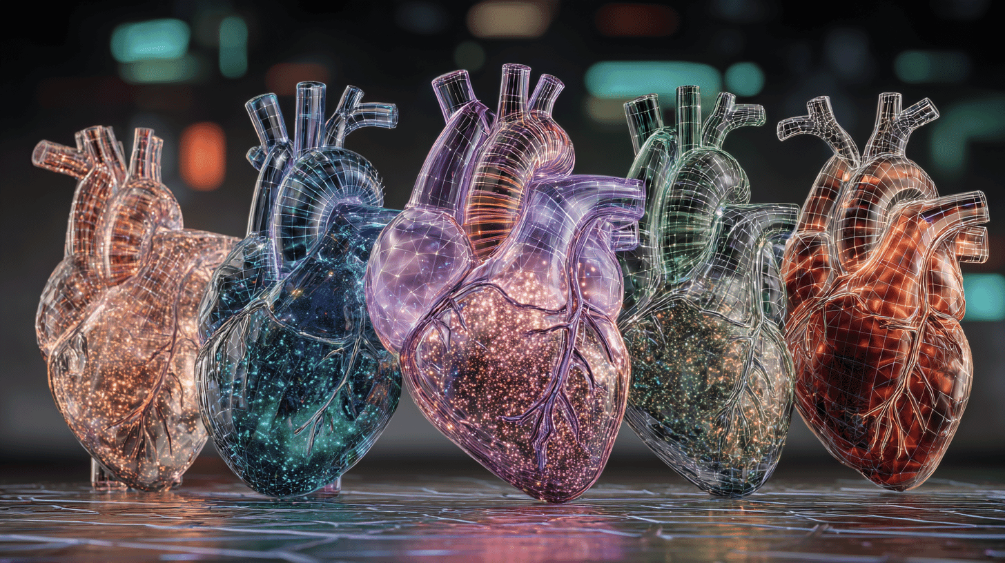
🔬 How many cells does a newborn mouse have? About 610 million, and a team at the University of Tokyo has mapped the exact 3D location of every single one. They've built the world's first whole-body cellular atlas at single-cell resolution, and it could fundamentally change how we diagnose disease and develop drugs.
Making the Body Transparent to See Every Cell
A research group led by Professor Hiroki R. Ueda at the University of Tokyo Graduate School of Medicine, with visiting researchers Shota Yoshida and Katsuhiko Matsumoto, has constructed the "CUBIC Organ/Body Atlas," a three-dimensional map that records every cell across all major organs of mice, and across the entire body of newborn mice, at single-cell resolution. The work was funded through JST's ERATO program and carried out with researchers from the Cancer Institute of JFCR, Juntendo University, Osaka University, Iwate Medical University, and Kurume University.
The paper was published online in Cell on February 25, 2026 US Eastern time, and selected for the cover of the March 19 issue.
Why It Matters: The Leap from 2D Slices to 3D Maps
For over a century, pathology and biology have run on 2D analysis: cut tissue into thin slices, examine under a microscope. The method has produced enormous medical advances, but it can only show a tiny cross-section. Rare cell populations, uneven disease patterns, and spatial relationships between tissue regions are easy to miss in a single thin slice.
Professor Ueda's team solved this by combining two key technologies: tissue clearing (making organs transparent) and advanced 3D imaging.
The CUBIC Method: Making Organs as Clear as Glass
CUBIC (Clear, Unobstructed Brain/Body Imaging Cocktails and Computational analysis) is a tissue-clearing technique originally developed at Japan's RIKEN institute in 2014. It works by removing lipids (fats) and pigments from biological tissue and adjusting the refractive index, essentially making organs literally transparent while preserving their 3D structure.
However, different organs require different treatment protocols. Kidneys need ten days for lipid removal, twice the usual duration, while livers require a gradual increase in the concentration of refractive-index-matching solutions. They optimized clearing protocols for all 11 major organ types in adult mice and adapted the technique for entire newborn mouse bodies.
A Custom Microscope for the Ultimate Challenge
To image these transparent specimens, the team developed a custom light-sheet microscope. Light-sheet microscopy works by scanning a thin sheet of light through a sample, but conventional systems struggle to combine high resolution with the ability to image large, thick specimens.
The team's new instrument can capture 3D images of organs and entire bodies spanning tens of millimeters, while maintaining resolution sharp enough to distinguish individual cells. This was the key engineering breakthrough that made the whole project possible.
Pinpointing 610 Million Cells in a Single Body
From the 3D images, the team extracted the position of every cell. In newborn mice, they found an average of approximately 610 million cells in males and 530 million in females. All of this positional data was compiled into a 3D atlas with a standardized coordinate system.
This coordinate system is what makes the atlas truly powerful. Just as GPS coordinates let anyone describe any location on Earth using the same system, the CUBIC atlas lets researchers overlay cell data from different animals, disease conditions, or drug treatments onto a common reference frame for direct comparison.
Real-World Applications: Mapping Immune Cells Across the Body
To demonstrate the atlas in use, the team mapped the distribution of macrophages, the immune cells that engulf foreign material, across the entire body.
The results revealed striking differences between organs. The spleen, for example, showed clusters of macrophages concentrated in specific zones, suggesting organ-specific immune roles. The team also tracked changes in cell distribution during kidney development, organ damage from drug administration, and shifts in immune cell populations during inflammation, all in 3D and quantitatively.
Where This Fits in Global Cell Atlas Efforts
The race to map cells is one of the biggest scientific endeavors of our time. The Human Cell Atlas, an international consortium launched in 2016 with more than 3,600 members across 102 countries, aims to create a complete reference map of every human cell type. As of 2024, approximately 62 million human cells had been mapped across 18 biological networks.
Spatial biology, the study of cells in their native tissue context, is among the fastest-growing fields in the life sciences.
Most existing cell atlases, however, rest on single-cell sequencing, which isolates cells from tissue and loses their positional information in the process. Spatial context is usually added back by probing thin sections with molecular markers, which covers only small regions at a time. What the CUBIC atlas supplies is the missing piece at whole-organ and whole-body scale.
Because the data is structural rather than molecular, it can be integrated with existing gene expression and spatial transcriptomic datasets, bridging shape information and molecular information.
The Road to 3D Pathology and Drug Discovery
Today, pathologists diagnose disease by examining thin, stained tissue sections on glass slides, much as their predecessors did a century ago. If the CUBIC approach is eventually applied to human tissue, entire organs could be examined in 3D: where a cancer has spread through an organ, how immune cells have infiltrated a tumor from every direction, how a drug's effects distribute unevenly across tissue regions. That is what "3D pathology" would mean in practice.
In drug development, whole-body mapping of effects and side effects could change how candidates are evaluated. The caveat is scale: this work was done in mice, and human organs differ in both size and composition. Clinical application is still some distance away.
The CUBIC Organ/Body Atlas is, in essence, Google Maps for the body. Just as a map organizes every location on Earth into a shared coordinate system that anyone can use, this atlas puts every cell in the body onto a shared 3D reference that researchers can lay their findings over.
How is cell atlas research progressing in your country? And what applications of 3D cellular mapping interest you most: disease diagnosis, drug development, or something else? We'd love to hear your perspective.
References
- https://www.jst.go.jp/pr/announce/20260226/index.html
- https://doi.org/10.1016/j.cell.2025.12.057
- https://www.humancellatlas.org/
- https://www.nature.com/articles/s41587-025-02699-5
- https://www.kurume-u.ac.jp/joint/topics/research/698abd4935283c1577d0addc/
- https://www.eurekalert.org/news-releases/1120896






Global Discussion
16 comments