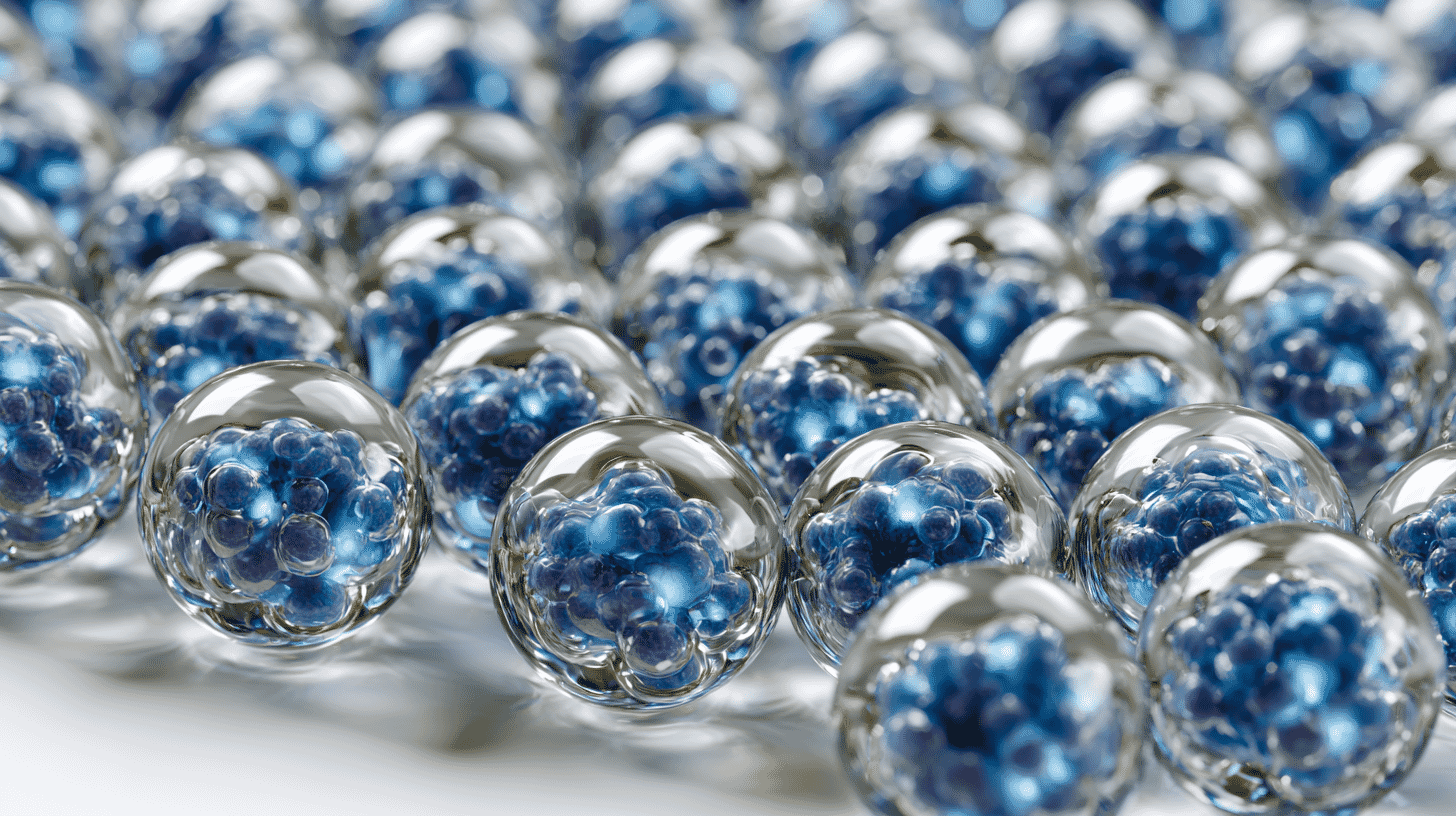
🧫 Imagine you want to test a new cancer drug. You need tens of thousands of tiny, near-identical capsules, each holding a few living cells, so your results are consistent enough to trust. Until now, making them took a delicate, expensive machine and months of training. A Japanese team has just shown you can do the same job with a desk-top shaker and a hot-and-cold water bath. It matters more than it sounds.
A bottleneck most people never hear about
When scientists test drugs or study how cells behave, they increasingly want cells grown in 3D rather than flat on a plastic dish. A flat layer of cells behaves differently from cells inside a living body. Cells clustered into a tiny ball, a "spheroid," behave much more realistically, which makes experiments more meaningful.
One reliable way to grow those clusters is to seal cells inside microscopic gel capsules, each smaller than a grain of sand. The capsule works like a private greenhouse: it holds the cells in place, shields them, and lets nutrients flow in.
The hard part is consistency. For an experiment to be trustworthy, every capsule has to be almost exactly the same size. A batch of wildly mismatched capsules produces messy data that's hard to compare. And for drug screening you don't need ten capsules; you need tens or hundreds of thousands of them.
For years, the standard way to make uniform capsules at that scale has been a microfluidic device: a chip threaded with hair-thin channels that drips out one identical droplet at a time. It works, but it is practically its own small craft. The chips are costly, they clog, they need a trained operator, and because they form capsules one droplet at a time, scaling to hundreds of thousands is slow. For a lab that simply wants to run an experiment, that is a real wall to climb.

Source: Japan Science and Technology Agency (JST) press release
How the ETE method works: borrowing a trick from jelly
The new method comes from Professor Sadao Ota at the University of Tokyo's Research Center for Advanced Science and Technology, working with Natsuko Otaki, Yuki Goda, and Eiryo Kawakami at Chiba University's Graduate School of Medicine and Kazuki Hattori of Kumamoto University. They call it Emulsion-Templated Gel Embedding, or ETE, and published it in the journal ACS Biomaterials Science & Engineering on May 21, 2026. Chiba University and the University of Tokyo have also filed a related patent application (PCT/JP2025/043191).
The clever idea is to let a pre-made bead do the precision work that the microfluidic machine used to do.
It runs roughly like this. First, the team prepares gelatin beads that are all the same size. Gelatin is the protein that sets a dessert like Jell-O: soft when warm, firm when cold. These uniform beads are mixed with the cells. A simple shake on a vortex mixer, a small and inexpensive piece of kit that sits on any lab bench, breaks the mixture into droplets. Because each droplet forms around one bead, the droplets all come out the same size. Warm the mix and the gelatin melts and engulfs the cell; cool it and the gelatin sets again, now with the cell sealed inside. The result is a uniform gelatin bead with a cell at its core.
Then the team repeats the trick. They use that cell-containing bead as a fresh template, wrapping it in a shell of agarose, a firmer gel and the same substance as the agar jelly used in Japanese cooking and in lab petri dishes. Finally they melt the gelatin out from the inside, leaving a hollow agarose capsule with the cell sitting in its chamber.
The whole process needs only a vortex mixer and the ability to raise and lower temperature. No microfluidic chip. And rather than dripping out capsules one at a time, a single round can produce them by the hundreds of thousands; the published work demonstrated more than 100,000 cell-containing capsules in one workflow, far faster than the chip-based route.
The real innovation: not a better capsule, but a lower wall
It's worth being precise about what is and isn't new here. ETE doesn't claim a fundamentally better capsule. Cells inside survive and multiply about as well as they do with the microfluidic method, and they form the same realistic 3D spheroids. The team also showed two useful extras: more than one type of cell can be packed into a single capsule, and the capsule can later be dissolved with an enzyme to recover the cells alive. The demonstrations centered on suspension cells, which float freely in culture; applicability to adherent cells is still being explored.
The breakthrough is access. ETE takes a capability that used to belong to labs with specialized hardware and specialized people, and puts it within reach of almost any biology lab with a basic bench. In the researchers' own framing, it lowers the barrier to entry for cell encapsulation, turning a niche technique into something closer to routine.
Scientific progress is often gated less by whether something is possible than by how many people can actually do it. When a method becomes cheap and simple, more labs try more ideas.
What it could unlock for drug discovery and regenerative medicine
Two areas stand to gain.
The first is drug discovery. Testing a candidate drug means exposing cells to it and watching what happens, ideally across thousands of nearly identical samples, so a real effect can be told apart from random noise. Uniform, mass-produced cell capsules suit that kind of large-scale screening well. Cheaper, faster capsule production means more compounds can be tested earlier, and the early stages are exactly where drug development tends to waste the most time and money.
The second is regenerative medicine and cell engineering. Encapsulated cells are studied as a way to shield transplanted cells from a patient's immune system, and as building blocks for lab-grown tissue. Producing large, uniform batches reliably is a basic requirement for any of that to scale. It also connects to a broader push, in Japan and abroad, to grow cells in conditions close enough to the human body that researchers can lean less on animal testing.
None of this is a finished product. ETE is a freshly published laboratory method, and turning a method into standard practice takes time. But the direction is clear: make the tool simpler, and you widen the door.
Japan's quiet bet vs. America's organoid gold rush
In the US, 3D cell culture has grown into a real industry. Organoids, tiny lab-grown clumps of tissue that mimic a real organ, sit at the center of a fast-expanding market: according to market research from Fortune Business Insights, the global human-organoid market was valued at roughly $1.18 billion in 2025 and is projected to reach about $5.7 billion by 2034. The same firm puts North America's share of that market at 41.5% in 2025. Companies there sell organoids, "organ-on-a-chip" devices, and screening services; Emulate, for one, built its business on sophisticated microfluidic chips. Regulation is pushing in the same direction. In April 2025 the FDA announced plans to phase down animal-testing requirements for certain drug classes, and on October 6, 2025 it cleared an Investigational New Drug application whose efficacy evidence came solely from a human vascularized tumor organoid model, developed by the San Diego company Qureator, with no animal proof-of-concept study. The drug was the cancer candidate BAL0891 in combination with a checkpoint inhibitor. That was permission to begin human trials, not approval of a finished drug.
That American model is powerful, but it is largely a model of selling advanced platforms and services: capability delivered as a product.
The Japanese contribution here is different in spirit. ETE isn't a platform to buy; it's a technique to adopt. It came out of public, government-funded research, backed by Japan's JST and AMED among others, and its explicit aim is to remove hardware rather than sell it, patent filing notwithstanding. If the American organoid boom is about building better machines, ETE is about making the machine unnecessary.
Neither approach is "right." A precision microfluidic platform still does things ETE cannot, especially the exquisite control needed to seal exactly one cell at a time. The two philosophies are complementary, and 3D cell culture probably needs both: the high-end industrialists and the great equalizers.
What happens next
The honest answer is that it depends on adoption. A method only matters if other labs pick it up, stress-test it, and find it holds up across many cell types and many pairs of hands. The published study is a strong proof of concept; the verdict comes from the labs that try it next.
But the appeal is easy to see. Cut the cost and skill a task requires, and you don't just help the researchers already doing it; you let in the people who were locked out before. That is often how a quiet methods paper ends up mattering more than a flashy one.
In your country, is cutting-edge biology concentrated in a handful of well-funded labs, or is there a push to put these tools within everyone's reach? When a technique gets cheaper and simpler, who do you think benefits most where you live?
References
- https://www.jst.go.jp/pr/announce/20260526-3/index.html
- https://www.jst.go.jp/pr/announce/20260526-3/pdf/20260526-3.pdf
- https://doi.org/10.1021/acsbiomaterials.5c02129
- https://www.genengnews.com/industry-news/top-10-organoid-companies/
- https://www.cbinsights.com/investor/hubrecht-organoid-technology








Global Discussion
3 comments