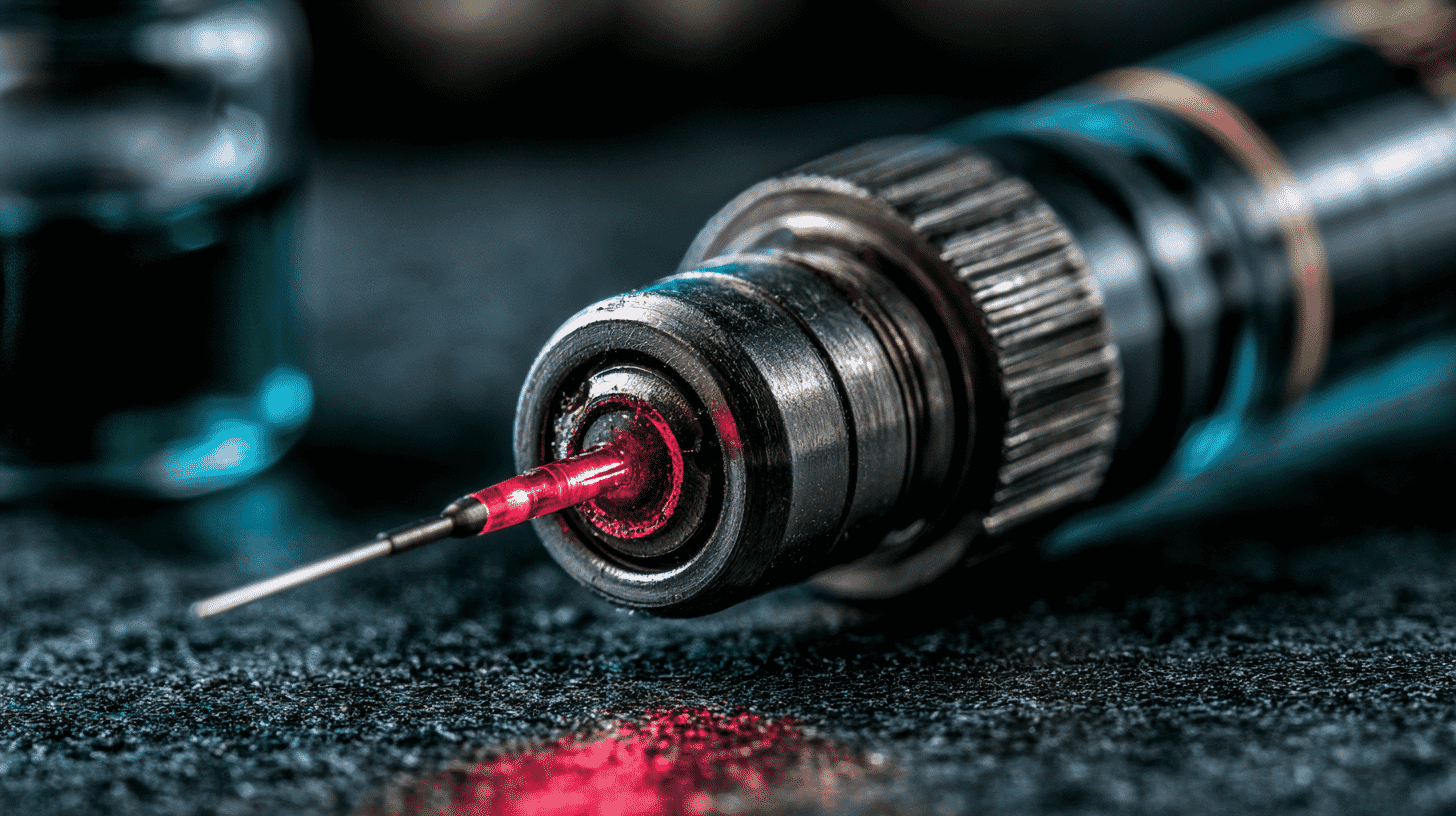
🔬 A laser fired from inside the tumor. A protein designed by an AI to slip past the immune system.
A team at Tohoku University has put those two ideas together, and in mice with colon cancer, one injection followed by one laser session made the tumors disappear within two weeks. The word carrying all the weight in that sentence is "mice." But the two problems the team went after are precisely the two that have kept this treatment idea parked in the laboratory for most of the past twenty years.
Killing cancer with heat is an old idea that keeps stalling
Photothermal therapy sounds almost crude when you spell it out. Inject particles that soak up near-infrared light and turn it into heat. Wait for them to collect in the tumor. Shine a laser. The tumor cooks.
Near-infrared light is the part that makes it plausible. Human tissue is relatively transparent at those wavelengths, which is why a flashlight pressed against your palm glows red on the other side. That transparency is what allows you to heat something buried under skin and muscle without heating everything in between.
The appeal is easy to see. Nothing gets cut open, no drug circulates through every organ, and no ionizing radiation is involved. Chemotherapy is essentially a poison you hope damages the tumor faster than it damages you. Radiation deposits energy along the whole beam path whether you want it there or not. Photothermal therapy, in principle, only does something where the particles are.
In practice, two things go wrong.
The first is that the body throws the particles away. Macrophages, the immune system's cleanup crew, read foreign objects in the bloodstream as garbage and haul them off to the liver and spleen. Not enough of the dose ever arrives.
The second is the light itself. Skin, fat and muscle absorb and scatter a laser beam, so reaching a deep tumor from outside the body takes more than 1 watt of power, and at that level you start burning the skin on the way in.
Source: Tohoku University press release
The raincoat problem
For decades, the answer to the first problem has been PEG, or polyethylene glycol, a petroleum-derived polymer wrapped around nanoparticles to make them look boring to the immune system. It appears in a great many approved drugs. It is also in the lipid nanoparticles of mRNA COVID vaccines.
PEG has two known weaknesses. Repeat doses can prompt the body to produce antibodies against it, after which subsequent doses get flushed from the blood unusually fast. Researchers call this accelerated blood clearance, and cancer treatment usually means repeat doses. Anti-PEG antibodies are detectable in a substantial share of the general population, and mRNA vaccination can raise their levels further, though how much this matters in real clinical settings is still being argued over. The second weakness is blunter: PEG does not break down inside the body.
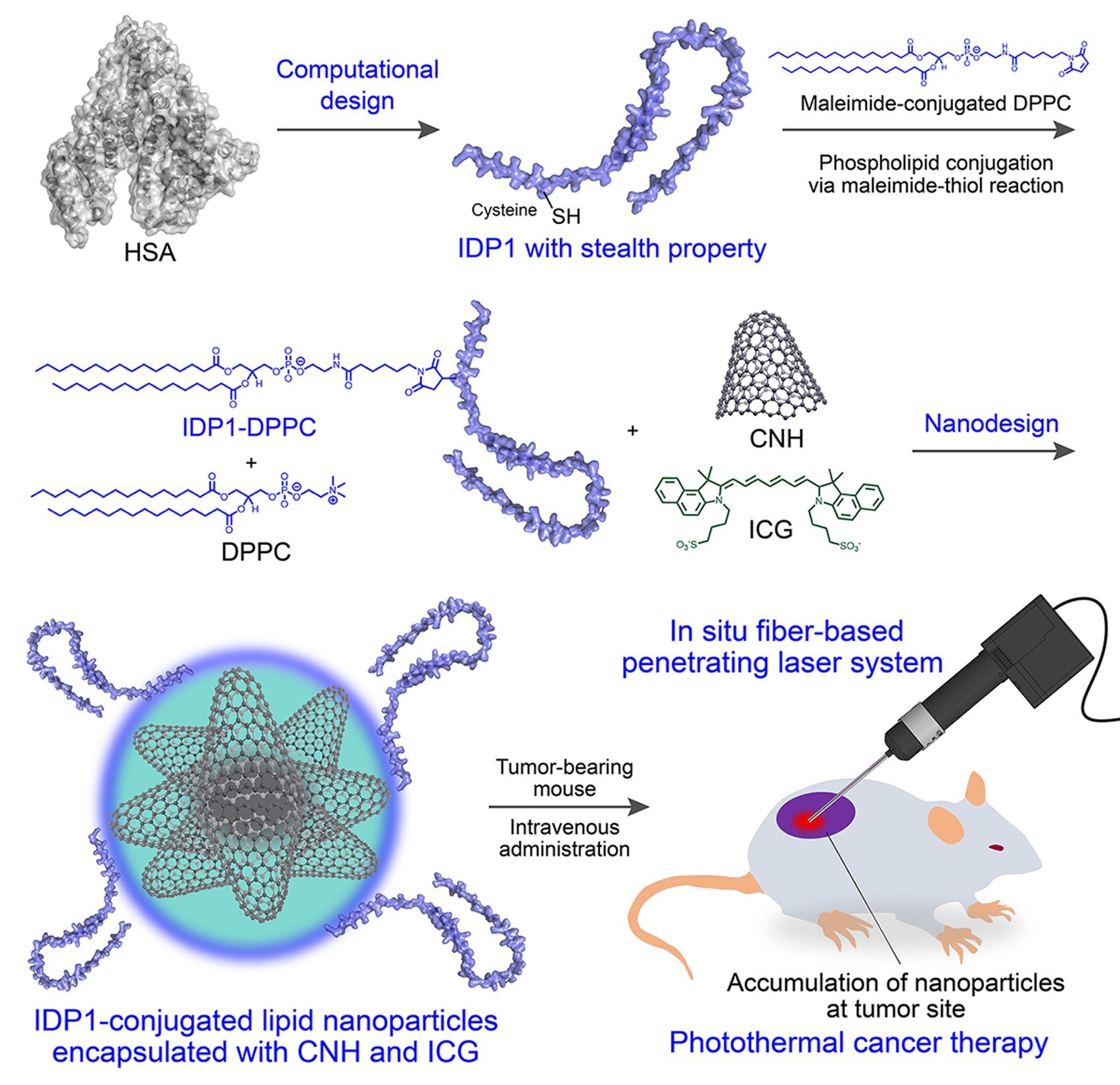
So Eijiro Miyako's group at Tohoku University's Institute of Multidisciplinary Research for Advanced Materials built a replacement. They started from human serum albumin, the most abundant protein in blood, roughly 200 grams of it in a 60 kg adult, and already good at going unnoticed by immune cells. They analyzed its structure, kept only the amino acids with the largest contact area with water, then ran the modified sequences through AlphaFold, the protein structure prediction AI from Google DeepMind, to find which stretches would refuse to fold into any fixed shape.
That refusal is the whole point. An intrinsically disordered protein flutters loosely in water instead of holding a rigid form, and that floppiness combined with a strong affinity for water is what makes it hard for immune cells to get a grip on. From the candidates, the team selected the one with the highest hydrophilicity and a slight negative charge and named it IDP1.
It runs about 100 amino acids, small enough to mass-produce in E. coli. In mice, nanoparticles coated with IDP1 stayed in circulation with a half-life of 150.5 minutes, against 17.8 minutes for PEG. And because IDP1 is a protein, the body's own enzymes eventually take it apart into harmless amino acids.
What it coats is a particle called IDP1-CNH/ICG, a lipid sphere roughly 200 nanometers across. Inside it are carbon nanohorns, a carbon material that converts light into heat efficiently, and indocyanine green, a fluorescent dye already approved by the FDA.
An endoscope that fits inside a needle
The light problem got a hardware answer, developed with Air Water Inc. and a group led by Hiromasa Yamashita, who also holds a project associate professorship at Teikyo University.
If the laser cannot get through skin, stop sending it through skin. Fiber systems that deliver light directly into tissue already exist, but they have been too thick for small-animal work and more invasive than the researchers wanted.
Air Water's contribution came out of its work on graded-index plastic optical fiber, which it adapted into a lens. The resulting rigid endoscope has a lens 0.5 mm across, small enough to sit inside the bore of a standard 16-gauge needle. Push the needle into the tumor and you can fire an 808 nm laser from within it.
The numbers make the case. Irradiating from outside the body at 700 mW raised the tumor temperature by about 40°C. Firing from inside at 500 mW, which is 28 percent less power, produced a rise of 36.3°C. Five minutes into irradiation, the inside of the tumor reached roughly 67.7°C.
The same device also carries a fluorescence imaging channel, exciting at 785 nm and detecting between 810 and 860 nm. That lets the operator watch where the indocyanine green has actually collected and aim accordingly, with one instrument instead of two.
In mice, the tumors vanished
In cell culture, IDP1-coated particles were taken up far less by RAW264.7 macrophages than uncoated ones. Under contact laser irradiation, Colon26 cancer cells died in proportion to particle concentration, while normal MRC5 cells were largely spared.
In live mice, 24 hours after intravenous injection, fluorescence at the tumor site substantially exceeded that in the liver, kidney, heart, spleen and lungs.
Then the treatment. Every mouse given IDP1-CNH/ICG plus contact irradiation lost its tumor within 14 days of a single laser session, with no recurrence across more than 40 days of observation. Body weight held steady and blood work came back normal.
The control groups matter as much as the treated one. Laser alone did not clear the tumors. Drug plus conventional external irradiation did not clear them either. The result depended on both halves working together.
The work appeared in Small Science on July 14, 2026, and was announced on July 21. As of July 2026, the team's stated next steps are non-clinical safety testing and research aimed at early clinical application. The endoscope is pointed at cancers it can physically reach: head and neck, esophageal, breast, colorectal.
None of which is a treatment yet. Five mice per group, a transplanted tumor model, 40 days of follow-up. Mouse tumors have been cured thousands of times over.
The United States has already tried this in humans
Nanospectra Biosciences, based in Houston, has been running the most clinically advanced version of this idea for years. Its AuroLase therapy uses gold-silica nanoshells about 150 nm across. They go in intravenously on the first day and the laser follows on the second, delivered through interstitial optical fibers placed through the perineum under MRI and ultrasound fusion guidance. A pilot study published in PNAS in 2019 enrolled 16 men with low to intermediate risk prostate cancer; 15 went through the two-day procedure, and 13 of those showed no detectable cancer at one year. The researchers were explicit that the trial was too small to measure efficacy formally.
So neither the nanoparticle nor the fiber inside the tissue is new. What the Tohoku work changes is the materials science at both ends. Gold-silica shells do not degrade in the body, a long-term safety question regulators take seriously, while carbon nanohorns inside a lipid capsule under a protein coat present a different profile. And shrinking the fiber to needle scale with imaging folded into the same instrument is a different engineering problem than placing multiple introducer trocars through a targeting grid.
The wider picture flatters no one. Thousands of photothermal therapy papers have been published. The number that have reached human trials is tiny. Particles that behave beautifully in a mouse tend to lose their uniformity once manufacturing scales up, and materials with heavy metals or non-degradable cores face a hard road through regulators. The distance between a mouse and a person is where this entire field has spent two decades.
Is heat ever going to be a real option
What this group has contributed is characteristic of a lot of Japanese materials research: not a new principle, but engineering that makes an old principle less impractical. An AI-designed protein to stand in for the plastic that has been the default for decades, and lens technology borrowed from a company that started out in industrial gases.
Whether that adds up to a treatment depends on results that do not exist yet. But the goal is not abstract. "Fewer side effects" means something specific to anyone who has watched a family member go through chemotherapy.
A university lab and a company that mostly makes something else: in Japan, that pairing is a standard shape for this kind of work. How does it look where you are? Is nanomedicine something people actually hear about, or does it stay locked inside universities?








Global Discussion
4 comments