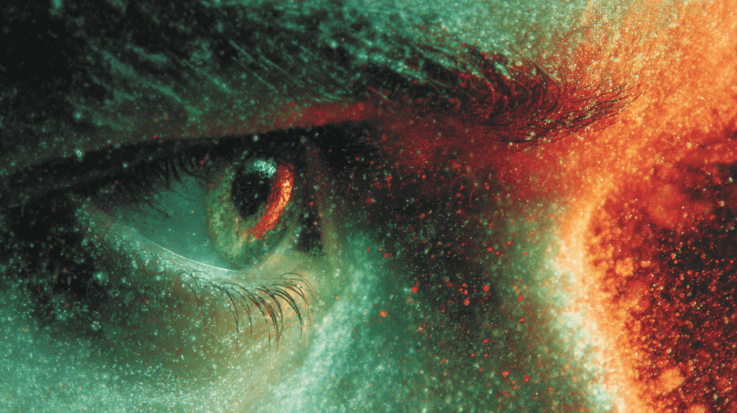
🔬 Two of the proteins inside your eye are nearly identical twins. Their genetic blueprints overlap by about 96 percent. Yet one lets you see red, and the other lets you see green. The whole difference between a ripe strawberry and an unripe one comes down to a gap of roughly 30 nanometers in the light they absorb, a distance thousands of times thinner than a hair. For decades, no one could explain, atom by atom, why that tiny gap exists. In June 2026, a team led from Nagoya froze the proteins still and finally looked inside.
The puzzle hiding in your retina
Daytime color vision runs on three light-sensing proteins packed into the cone cells of the retina. They are tuned to long, medium, and short wavelengths, which we experience roughly as red, green, and blue. The red pigment responds best to light around 560 nanometers, the green one around 530. That 30-nanometer offset is the hinge your whole sense of "red versus green" swings on.
Here is the strange part. The red and green pigments are close evolutionary cousins, born from a gene that duplicated relatively recently in primate history. Their amino acid sequences are about 96 percent identical. A handful of differences, no more, separates seeing a stop sign from seeing the leaves behind it.
When this system is even slightly miswired, the result is red-green color vision deficiency, the most common form of color blindness by far, making up roughly 99 percent of all cases. It affects about 1 in 12 men of European descent, around 5 percent of Japanese men, and far fewer women, because the relevant genes sit on the X chromosome. For all of that, the structural reason behind the 30-nanometer gap stayed a mystery. Researchers knew which amino acids differed. They could not see how those differences, arranged in three-dimensional space, actually bent the proteins' response to light.
A protein that fell apart before it could be seen
Part of the problem was that the cone pigments are delicate. The structure of rhodopsin, the pigment that handles dim night vision in our rod cells, was solved more than twenty years ago. The cone pigments resisted. They are unstable outside their natural setting, awkward to produce in usable amounts, and prone to losing their shape during the very experiments meant to capture them.
So the cause of the most common color difference in human vision sat just out of reach, the target dissolving before anyone could photograph it.
Frozen at the moment before light
The breakthrough came from cooling the problem down, literally. Using cryo-electron microscopy and single-particle analysis, the group, led by Kota Katayama and Hideki Kandori at the Nagoya Institute of Technology and joined by researchers from the University of Tokyo, Kyoto University, and Italy's University of Siena, determined the three-dimensional structures of the red and green cone pigments from a macaque, a primate whose color vision closely mirrors our own. Crucially, they captured the pigments in their dark state, the resting shape they hold before any light arrives. That resting state is where the color tuning is set, and it had never been seen at atomic resolution for the red and green pigments before. The team then cross-checked the picture against heavy quantum-chemistry calculations, so the structure and the physics agreed. The results went online in Science on June 25, US Eastern time.
The timing was striking. The Nagoya-led paper was one of three the journal published that same day, a cluster it billed as cracking color vision. A Switzerland-led group reported the dark-state structures of the human blue and green pigments, and a China-led group reported all three human pigments in their activated state. Three teams came at the problem from three different angles, all at once. The distinctive target of the Japan-led work was the oldest question in the set: why red and green, the near-twins, part ways.
The answer was three amino acids
When the structure finally came into focus, the answer was almost suspiciously small.
The molecule that actually catches light, called retinal, is essentially the same in both pigments. What differs is the protein pocket cradling it. Three water-friendly amino acids, unique to the red pigment, sit in slightly different positions and carry slightly different electrical character, what chemists call a dipole. Together they build a particular electrostatic environment around the light-catching molecule, and that environment nudges which wavelength it prefers. One of the three, a threonine, contributes the most; the others fine-tune. It is not a hard mechanical shove that separates red from green. It is a faint electrical tilt.

Source: JST / Nagoya Institute of Technology press release
A side door that was already open
The structure also gave up a bonus the team was not necessarily hunting for.
When they compared the cone pigments to rhodopsin, they noticed openings along the side of the protein that faces the cell membrane, openings rhodopsin's inactive form does not have. It turns out the light-catching retinal exits and enters through different routes. The exit passage forms when the pigment is activated, much as it does in rhodopsin. But in the cone pigments, the likely entrance passage appears to be open already while the protein sits in the dark, waiting.
That detail matters more than it sounds. An entrance left ajar means spent retinal can be swapped for fresh retinal quickly, so the pigment recharges fast. The cone pigments also hold a looser, almost ready-to-fire shape even in the dark. Add it up, and you get exactly what daytime vision demands: cones that fire continuously in bright light and reset before the next photon, keeping color steady as the world rushes past.
What comes after the 30-nanometer answer
For now, the practical payoff sits in the future tense. Hereditary red-green color vision deficiency arises from changes in these very pigments, and a precise atomic map is the kind of thing drug designers and gene-therapy researchers have wanted for a long time. The researchers, and the institutions announcing the work, are careful to frame medical applications as expected rather than delivered.
But there is a subtler idea tucked inside the finding. If three amino acids and a faint electrical tilt set where "red" begins, then the border between red and green stops being a fixed line in nature and becomes a setting, written slightly differently in different proteins, and in different people.
So the next time you and someone else disagree about whether a shade is green or teal, consider that you might both be right, each reading the world through your own pocket of three amino acids. Is the red you see the same red your neighbor sees? And in your country, how much thought goes into color blindness, in classrooms, on road signs, in everyday design?





Global Discussion
4 comments