
Repeated IVF cycles that never end in pregnancy are known as implantation failure, and one suspected cause is the uterus's own constant, subtle motion. A team at the University of Tokyo has built an AI model that reads roughly 18 seconds of that motion, captured by MRI, and uses it to predict whether pregnancy will follow. The point is to turn something doctors used to judge by eye into a number.
When Good Embryos Won't Implant
In vitro fertilization (IVF) has given hope to millions of couples worldwide, yet its success rate remains stubbornly limited. Japan is one of the world's heaviest users of the technology: the Japan Society of Obstetrics and Gynecology counted 543,630 assisted reproductive technology (ART) treatment cycles in 2022. Even so, success rates fall to around 25% for patients in their early forties.
The most frustrating case is recurrent implantation failure (RIF): embryos that look healthy and viable are transferred, and pregnancy never takes hold. It is like planting good seed in soil that, for reasons nobody can pin down, keeps rejecting it.
Scientists have long suspected that uterine peristalsis, the subtle wave-like contractions the uterus constantly performs, plays a critical role. These contractions normally quieten during the implantation window; when they stay excessive or overly strong, they may physically prevent an embryo from settling in. Quantifying them has been the hard part. Until now, assessment came down to individual doctors watching MRI footage and forming a judgment.
Compressing Motion Into a Single Image
Researchers from the University of Tokyo Graduate School of Medicine and SIOS Technology Inc. set out to change this. Their study, published in Reproductive Medicine and Biology in March 2026, analyzed data from 188 patients diagnosed with RIF.
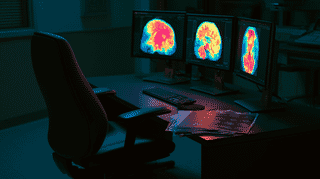
The team used cine MRI, a type of MRI that captures continuous images, essentially creating a video of the uterus in motion. The critical innovation was a proprietary method that converts four consecutive grayscale frames into a single RGB image, condensing roughly 18 seconds of uterine movement into one composite picture. It amounts to a motion signature of the uterus, captured in one frame that a deep neural network (DNN) can process efficiently.
That solved a practical problem. Feeding raw video into a deep learning system demands enormous compute and far more training data than 188 cases can supply. Distilling the time dimension into one image made the analysis feasible at that scale.
The DNN's output was then summarized into five statistical measures, which were combined with clinical information (such as age) in a Random Forest machine learning model to generate final pregnancy predictions.
The Numbers Tell the Story
The integrated model, combining cine MRI analysis with clinical data, achieved an area under the ROC curve (AUC) of 0.835, with accuracy of 75.4%, sensitivity of 87.9% and specificity of 58.3%.
The model using clinical data alone managed an AUC of only 0.617, with accuracy of 59.6%, sensitivity of 69.7% and specificity of 45.8%. Every one of those numbers improved once uterine motion was added.
To put this in perspective: the AI model incorporating uterine movement data correctly identified roughly 88 out of every 100 patients who would go on to become pregnant. Adding information about how the uterus actually moves transformed a marginally useful prediction tool into one with genuine clinical potential.
Why This Matters Beyond the Lab
The significance of this research extends in several directions.
First, it objectifies what was previously subjective. Doctors' assessments of uterine movement have always varied depending on experience and interpretation. This model provides standardized, reproducible metrics, a prerequisite for any diagnostic tool that hopes to be adopted widely.
Second, it shifts attention to the "receiving end" of implantation. Most AI work in fertility treatment has focused on grading embryo quality, which is judging the seed. This research shows the soil matters just as much, and that uterine function can be quantified and fed into a model.
Third, it opens the door to personalized fertility treatment. If a patient's movement pattern points to a low chance of implantation, clinicians might intervene before transfer, whether by calming excessive contractions with medication or by timing the transfer more precisely.
What Comes Next
The research team plans to test the model against an independent external dataset and add cases, to generalize it and refine its performance. Future work aims to fold in other uterine conditions, such as fibroids and adenomyosis, building toward a broader assessment of uterine health.
The limitations are real. This was a retrospective study: the model was developed and tested on historical data. Prospective trials, where the model guides live treatment decisions, are still needed. Additionally, cine MRI is not standard equipment at most fertility clinics, which could limit near-term practical application.
Nevertheless, the approach points toward reproductive medicine in which treatment decisions rest not only on a patient's age and history but on how their own uterus actually behaves.
That matters most in a country like Japan. In 2022 the government brought fertility treatment under public insurance, and that year 77,206 babies were born through ART, roughly one in every ten births. The more people go through treatment, the more valuable a tool that can explain why implantation fails becomes.
What role does AI play in fertility treatment in your country? How do you feel about using technology to predict and improve pregnancy outcomes? We'd love to hear your perspective!








Global Discussion
12 comments