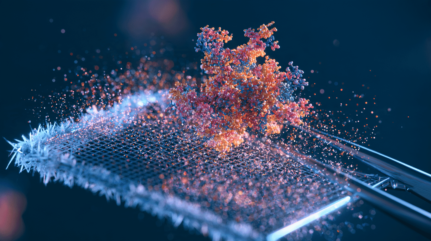
🔬 In a single overnight run, a microscope in northern Kyoto shoots 5,000 to 6,000 images of frozen proteins. Each image holds a few hundred particles. Assume 400 per image and that comes to about 2 million molecules in one night. This autumn, third-year undergraduates at Kyoto Sangyo University are learning to turn those particles into a 3D shape they can spin around on their own laptops.
The machine is a cryo-electron microscope, or cryo-EM, and a single one can cost millions of dollars.
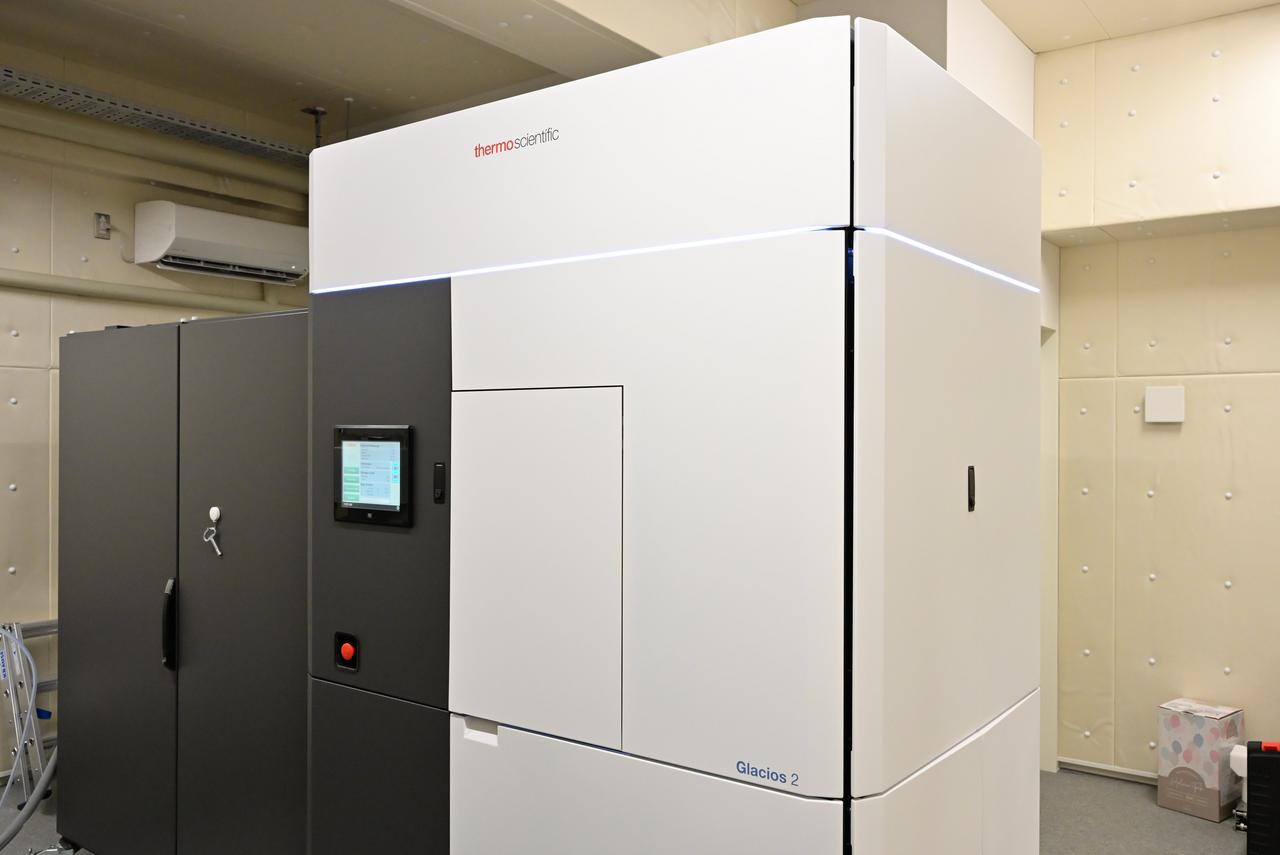
Source: Kyoto Sangyo University
Freeze first, photograph later
Electron microscopes can make out things far too small for ordinary light. For biologists, though, there was a catch: they were long believed useful only for dead matter. The electron beam wrecks biological samples, and the inside of the microscope is a vacuum, where water evaporates and molecules collapse.
Three ideas fixed that, and the people behind them shared the 2017 Nobel Prize in Chemistry. In the early 1980s, Jacques Dubochet found a way to cool water so fast that it turns into a kind of glass instead of ice crystals, so proteins keep their native form even inside the vacuum. Between 1975 and 1986, Joachim Frank developed an image-processing method that merges fuzzy 2D pictures into a sharp 3D structure. And in 1990, Richard Henderson used an electron microscope to build an atomic-resolution 3D model of a protein, proving the whole idea could work. According to the Nobel Prize announcement, the technique reached the atomic resolution researchers wanted in 2013.
You can't take one crisp photo of a single protein, because a beam strong enough for a sharp picture would destroy it. So you take an enormous number of faint snapshots of identical copies frozen at random angles, sort them by angle, and let a computer stack them until a clear 3D model emerges. Think of rebuilding a face from thousands of grainy security-camera frames.
Why scientists stopped growing crystals
Before cryo-EM, the standard way to see a protein's shape was X-ray crystallography, which has been developing since the 1950s. The catch is that you first have to coax the protein into a crystal, with its molecules lined up as neatly as grains of salt. There is no recipe for that. Researchers try thousands of conditions, one at a time.
Dohyun Im, an associate professor at Kyoto Sangyo University who teaches the new course, knows the pain. He told University Journal Online that he once spent three and a half years on a membrane protein that never crystallized. Cryo-EM skips the crystal entirely: freeze the sample and look. Im says structures can now appear within a year of planning an experiment, a pace he calls incomparable. He adds that X-ray crystallography often still gives finer detail, so labs pick whichever method suits the molecule.
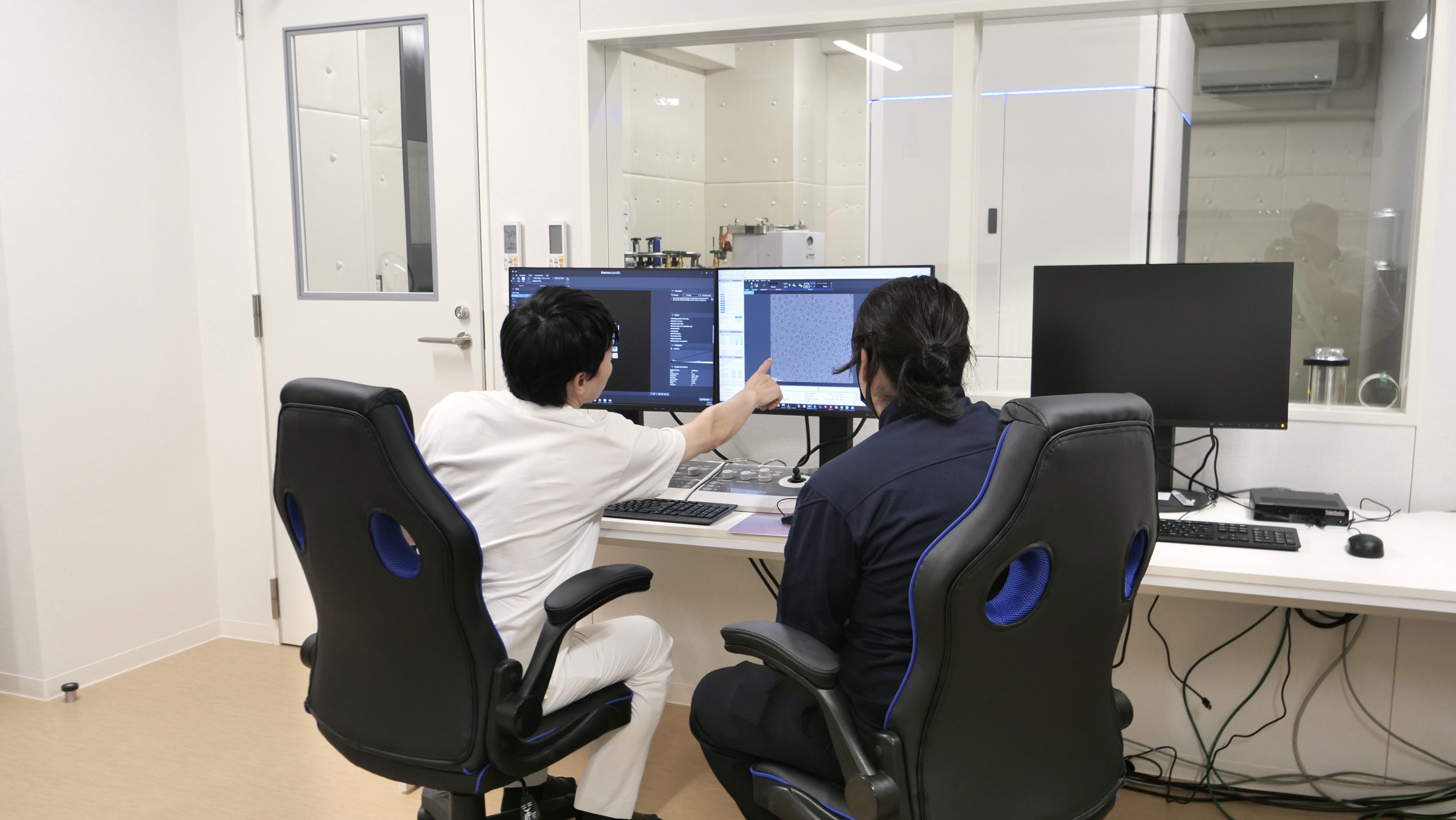
Source: Kyoto Sangyo University
What seeing a molecule lets you do
Drug discovery is where it is used most today, Im says. Many medicines work by slotting into a receptor on the surface of a cell, the classic lock-and-key picture. One large family of locks, G protein-coupled receptors (GPCRs), is said to be the target of about 30% of the drugs we take. For a long time nobody could see the shape of those keyholes. Now that they can, designing a key that fits has become easier. The fit is fussy, Im notes: a tiny change in a drug can make it hundreds of times stronger or weaker, or even switch the signal the receptor sends.
In early 2020, a team at the University of Texas at Austin and the US National Institutes of Health received the genome of the new coronavirus. Two weeks later they had engineered and made samples of its spike protein, the part that latches onto human cells. About 12 days later they had a 3D atomic-scale map and a paper submitted to Science, which published it on February 19, 2020. The university said the map would help researchers around the world develop vaccines and antiviral drugs.
When Henderson visited Kyoto Sangyo University for an international symposium on July 2, 2026, a student asked him what the most important research target would be. According to University Journal Online, he named the brain. Knowing the shapes of ion channels and GPCRs still doesn't explain how they add up to memory and learning, and he estimated that working it out would take another 30 to 40 years.
So, is it expensive? Very
McGill University in Canada told Nature Index in 2021 that buying and later upgrading its top-end Titan Krios cost 10 million Canadian dollars (US$8 million). It spent another 4 million Canadian dollars renovating a building to fit the 4.5-metre-tall instrument, and running it costs about 500,000 Canadian dollars a year, staff salaries included.
Smaller models are cheaper, not cheap. In 2020, a UK government contract to buy a Glacios, an earlier version of the model line Kyoto Sangyo installed, was valued at £1.4 million (about $1.85 million). Kyoto Sangyo did not give a price in its announcements.
In December 2020, the business daily Nikkan Kogyo Shimbun reported that 17 cryo-EMs were then available to researchers through a national support program, far behind the US and China. The education ministry had budgeted ¥3.2 billion (about $20 million) to install six more and to develop automated, remote-operation technology with companies.
As of October 2026, Kyoto Sangyo rents out its microscope by the day, counted as 24 hours from 10 a.m. Researchers from universities and public institutes, who are generally expected to publish their results, pay ¥55,000 (about $350) to run it themselves or ¥77,000 (about $490) with staff support. Companies, which generally keep their results private, pay ¥286,000 (about $1,800) or ¥495,000 (about $3,100). First-time users need a ¥33,000 (about $210) training session, and even the tiny metal grids the samples sit on cost ¥2,500 to ¥7,000 ($16 to $44) apiece, billed on top of the daily fee. University Journal Online notes that a day of measurement is said to cost close to ¥100,000 (about $630), even for academic use. (Dollar figures use the October 9, 2026 rates of ¥158.3 and £0.756 to the dollar.)
Why bring it into an undergraduate class?
Kyoto Sangyo, a private university, installed a Thermo Fisher Glacios 2 in autumn 2025, which by its own survey made it the first private university in western Japan to own a cryo-EM. The university says its researchers have published 10 cryo-EM papers since 2019 in Nature, Science and their sister journals, the most of any private university in Japan by its own count. Toshiya Endo, director of its Institute for Protein Dynamics, said his team used to travel to other universities and institutes to use such a machine, and that having one at home would change both the speed and the quality of their work.
Instead of keeping it for professors, the university built a course around it. Structural Cell Biology, a course for third-year students offered from autumn 2026, runs for 15 weekly sessions. The first half covers how a protein's shape relates to its job. In the second half, students watch a measurement, then do the processing themselves: cutting individual particles out of the images, grouping them by angle, averaging them into 2D views and finally building a 3D model. The university says hands-on cryo-EM analysis at the undergraduate level has no precedent in Japan. Beginner training does exist elsewhere, such as the free single-particle analysis workshops run by Osaka University's Institute for Protein Research, but as two-day workshops rather than a regular course.
Otsuka Pharmaceutical had hired Im as a crystallography specialist, and then he watched cryo-EM improve so fast that, as he puts it, he panicked. He quit, went back to Kyoto University and learned the technique from scratch. In 2024 he was first author of a study revealing the structure of VMAT2, a transporter that loads neurotransmitters into tiny sacs called vesicles.
He says he still remembers the thrill of first seeing the shape of the protein he had been chasing, and that moment is a big part of why he stayed in research. He wants students to feel it before they even join a lab, and thinks the experience could help them land jobs at drug companies.
Would a university near you put data from a machine like this in front of third-year students, or is top-end equipment kept for senior researchers? Tell us how it works where you are.
References
- https://univ-journal.jp/column/20261002255/
- https://www.kyoto-su.ac.jp/wr-news/20250425_850_cryo.html
- https://www.kyoto-su.ac.jp/research/kikou_pr/cryo/index.html
- https://www.nobelprize.org/prizes/chemistry/2017/press-release/
- https://news.utexas.edu/2020/02/19/breakthrough-in-coronavirus-research-results-in-new-map-to-support-vaccine-design/
- https://www.nature.com/nature-index/news/must-have-multimillion-dollar-microscopy-machine-cryo-em
- https://www.contractsfinder.service.gov.uk/Notice/17e288e2-2323-4799-96c9-7ec9f18a4d79
- https://newswitch.jp/p/25137
- https://doi.org/10.1038/s41467-024-51960-z
- https://www.protein.osaka-u.ac.jp/wp-content/uploads/2026/08/2026%E3%82%AF%E3%83%A9%E3%82%A4%E3%82%AA.pdf




Global Discussion
3 comments